Silica (SiO₂) Sensor Surfaces in Multi-Parametric SPR: Physicochemical Foundations and Biointerfacial Applications
Silica (SiO₂) surfaces are widely employed in label-free biosensing due to their well-characterized surface chemistry, physicochemical stability, and compatibility with a broad range of biomolecular systems. The surface is terminated by silanol (Si–OH) groups, which provide a versatile platform for chemical functionalization via silanization and related chemistries. These groups also confer a well-defined, pH-dependent interfacial charge, enabling the investigation of electrostatically driven adsorption processes. Due to these unique properties, the controlled study of biomolecule and polymer adsorption, as well as interfacial diffusion and mobility phenomena, can be easily monitored. Classical adsorption studies have demonstrated that silica can interact with a wide variety of natural and synthetic macromolecules through hydrogen bonding, electrostatic interactions, van der Waals forces, and specific interactions involving silanol functionalities. These properties render silica an ideal oxide surface for investigating interfacial phenomena relevant to biological and soft-matter systems.
Particularly, the study of biological membranes is important to understand their structure and assembly mechanisms in physiological environments and the monitoring of biophysical parameters such as bending and stretching rigidity, lipid composition and phase state, e.g. fluid or gel, can help predict mechanisms of interaction between target proteins or receptors within the membrane and ligands.
The integration of silica sensors with Multi-Parametric Surface Plasmon Resonance (MP-SPR) further enhances analytical capabilities. Unlike conventional single-wavelength SPR, MP-SPR enables simultaneous determination of multiple physicochemical parameters of the adsorbed layer, including thickness, refractive index, and surface mass density. This approach allows improved analysis of structural and compositional changes within soft and hydrated films and membrane lipid rearrangements, which are often challenging to characterize using traditional SPR.
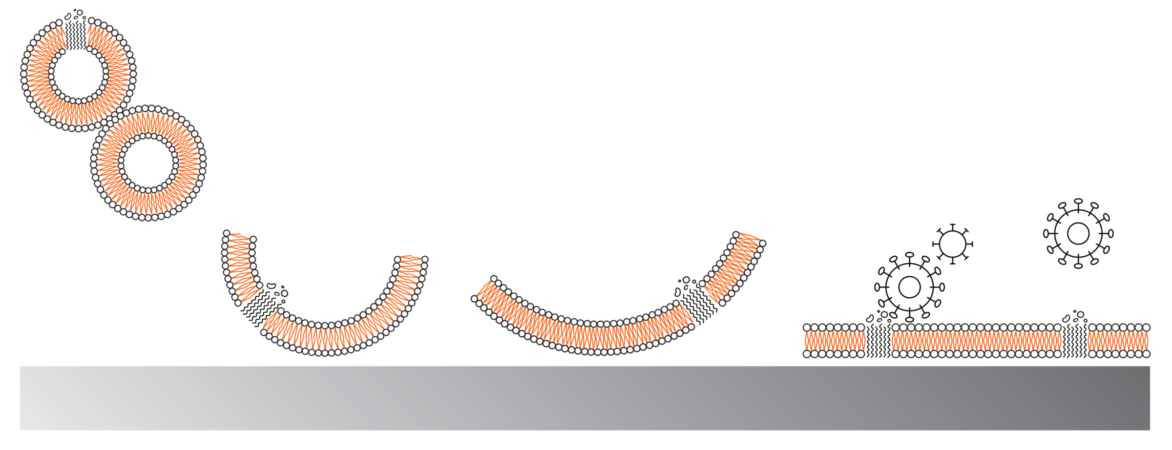
Norling et al. (2022) [1] illustrated the utility of silica sensors in membrane research. In their study, fluid-phase and gel-phase liposomes exhibited deformation dependent on ligand density within a supported lipid bilayer (SLB) formed on a silica-coated sensor. The silica surface used as a well-defined support for membrane-mimetic assays and as a reference interface for assessing nonspecific, was initially primed and activated with the buffer utilized for the experiments; successively, the liposome suspension was injected into the flow and the SLB is then formed after the liposomes rupture and unfold on the surface. Importantly, accurate structuring of lipid membranes to reproduce physiological conditions is essential for meaningful interaction studies, and the SLB formation could be accurately monitored by MP-SPR signal, first increasing after the injection of liposomes, then decreasing and reaching a plateau phase while the SLB forms. In this context, silica provides an optimal substrate to preserve bilayer integrity and lateral mobility, thereby enabling a closer approximation to the native membrane state [2].
A representative example is provided by Kubiak-Ossowska et al. (2017) [3], who investigated the adsorption of bovine serum albumin (BSA) onto silica sensors. Using MP-SPR, the authors monitored pH-dependent monolayer formation mechanisms and quantified the adsorbed mass, demonstrating the sensitivity of silica-supported SPR measurements to protein orientation and interfacial charge effects.
Similarly, Pinto et al. (2017) [4] demonstrated the electrostatically driven layer-by-layer deposition of chitosan and carbon dots (CDs) on silica surfaces. Real-time MP-SPR measurements enabled monitoring of multilayer growth under varying polymer and nanoparticle concentrations and pH conditions, while quantitative analysis of thickness and refractive index provided insight into the structural evolution of the assembled films.
In summary, the versatility of silica sensors in MP-SPR studies arises from their chemically well-defined and readily functionalizable surfaces. Slica supports the investigation of soft, hydrated, and swelling interfacial layers, where MP-SPR’s multiparametric modeling significantly improves characterization in terms of thickness and refractive index contributions. Importantly, silica surfaces provide an ideal platform for the formation of supported lipid bilayers, enabling the study of membrane structure, mechanical properties, and biomolecular interactions under controlled and physiologically relevant conditions. Moreover, silane-based modification strategies allow controlled tuning of surface charge and chemical functionality as a function of pH and ionic strength, facilitating systematic studies of electrostatic adsorption involving proteins, dendrimers, polymers, and supramolecular complexes. Finally, the technique enables real-time kinetic analysis and direct comparison with computational approaches, such as molecular dynamics simulations, thereby strengthening mechanistic interpretation of adsorption and structural phenomena at biointerfaces.
References:
[1] Norling, K., Sjöberg, M., Bally, M., Zhdanov, V.P., Parveen, N., Höök, F. (2022) Dissimilar Deformation of Fluid- and Gel-Phase Liposomes upon Multivalent Interaction with Cell Membrane Mimics Revealed Using Dual-Wavelength Surface Plasmon Resonance. Langmuir, 38 (8), 2550-2560 DOI: 10.1021/acs.langmuir.1c03096
[2] Ulmefors H., Nissa, J., Pace, H., Wahlsten,O. , Gunnarsson, A., Simon, D. T., Berggren, M., and Höök, F. (2021). Formation of Supported Lipid Bilayers Derived from Vesicles of Various Compositional Complexity on Conducting Polymer/Silica Substrates, Langmuir 37 (18), 5494-5505,
DOI: 10.1021/acs.langmuir.1c00175
[4] Tarciane da S. Pinto, Larissa A. Alves, Gabriele de Azevedo Cardozo, Victor H.O. Munhoz, Rodrigo M. Verly, Fabiano V. Pereira, João P. de Mesquita (2017). Layer-by-layer self-assembly for carbon dots/chitosan-based multilayer: Morphology, thickness and molecular interactions, Materials Chemistry and Physics, 186, 81-89, https://doi.org/10.1016/j.matchemphys.2016.10.032.
[3] Kubiak-Ossowska, K., Tokarczyk, K., Jachimska, B., & Mulheran, P. A. (2017). Bovine Serum Albumin Adsorption at a Silica Surface Explored by Simulation and Experiment. The Journal of Physical Chemistry B, 121(16), 3975–3986. https://doi.org/10.1021/acs.jpcb.7b01637
More peer-reviewed publications in which silica sensors were utilized with MP-SPR:
- Mateos H, Valentini A, Bellini T, Ambrosetti E, Bobba F, Stellacci F, et al. Surfactant Interactions with Protein-Coated Surfaces: Comparison between Colloidal and Macroscopically Flat Surfaces. Biomimetics. 2020;5(3):31. doi:10.3390/biomimetics5030031.
- Parkkila P, Viitala T. Partitioning of Catechol Derivatives in Lipid Membranes: Implications for Substrate Specificity to Catechol-O-methyltransferase. ACS Chem Neurosci. 2020;11(6):969–978. doi:10.1021/acschemneuro.0c00049.
- Fragasso A, de Vries HW, Andersson J, van der Sluis EO, van der Giessen E, Dahlin A, et al. A designer FG-Nup that reconstitutes the selective transport barrier of the nuclear pore complex. Nat Commun. 2021;12(1):2010. doi:10.1038/s41467-021-22293-y.
- Grad P, Edwards K, Hernández VA. Adhesion and Structural Changes of PEGylated Lipid Nanocarriers on Silica Surfaces. Physchem. 2021;1(2):133–151. doi:10.3390/physchem1020009.